Image
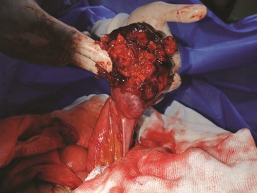
Jejunal gastrointestinal stromal tumour revealed by massive haematemesis (April 2023 [2])
A 56-year-old male experienced massive haematemesis for 2 days. Neither oesophagogastroduodenal endoscopy nor colonoscopy demonstrated a cause. An urgent abdominal computed tomography scan showed active jejunal bleeding. At explorative laparotomy, a large jejunal GIST was found.
View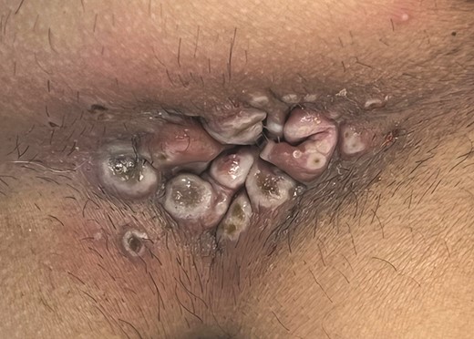
Image
Peri-anal monkeypox (April 2023 [1])
A 44-year-old male, who has sex with men, presented with proctalgia and peri-anal lesions. In addition, he had inguinal lymphadenopathy and ulcerated papules on his extremities, trunk, and face. On polymerase chain reaction testing, a diagnosis of monkeypox was made. Given the current prevalence, monkeypox should be considered in patients with new peri-anal lesions.
View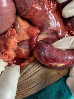
Image
Curious cause of small bowel obstruction (March 2023 [2])
A 60-year-old woman presented with abdominal pain, distension, and constipation. A computed tomography scan showed small bowel obstruction. At laparotomy, the small bowel was found to be obstructed by a fallopian tube. The fallopian tube band was released and right-sided salpingectomy performed.
View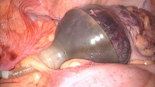
Image
Unusual cause of caecal ischaemia (March 2023 [1])
A 69-year-old man presented with right lower quadrant abdominal pain and fever. His medical history was notable for erectile dysfunction treated with an inflatable penile prosthesis. A computed tomography scan showed caecal ischaemia. Diagnostic laparoscopy (Video File) found the caecum had lodged in the empty reservoir of the penile prosthesis, causing ischaemia.
View
Image
Disseminated peritoneal leiomyomatosis (February 2023 [2])
A 55-year-old woman with a prior history of laparoscopic uterine myomectomy presented with an incisional hernia. Preoperative computed tomography reported calcified lymph nodes in the mesentery. At laparotomy, multiple tumours of up to 8 cm were found. An ileocaecal resection and omentectomy were performed. Following histopathological assessment, a diagnosis of Peritoneal Leiomyomatosis was made.
View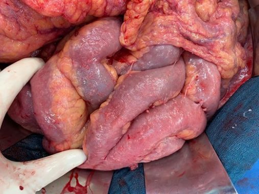
Image
Miliary peritoneal tuberculosis causing abdominal pain and ascites (February 2023 [1])
A 73-year-old male presented with abdominal pain and progressive ascites. Cytological assessment of the ascites found no abnormality. An exploratory laparoscopy was performed, which was converted when miliary seeding was identified. Histopathological examination demonstrated peritoneal tuberculosis.
View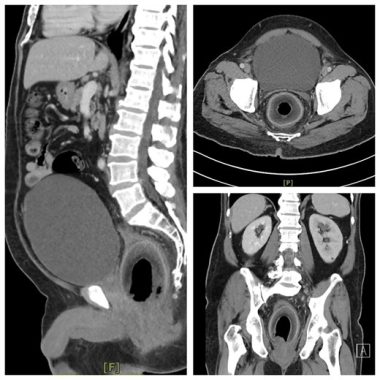
Image
Case of coconutention (January 2023 [2])
A 56-year-old man presented with a 2-day history of severe abdominal pain, anuria, and absolute constipation. Abdominal CT showed a large foreign body in the rectum, which compressed the prostatic urethra, causing urinary retention. A coconut was removed from the rectum via exploratory laparotomy.
View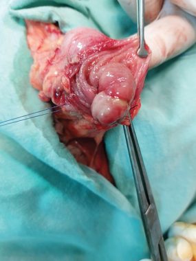
Image
Colonic lipoma (January 2023 [1])
A 46-year-old woman presented with abdominal pain. A computed tomography scan found colonic intussusception. She underwent a laparoscopic colectomy at which a giant lipoma was found.
View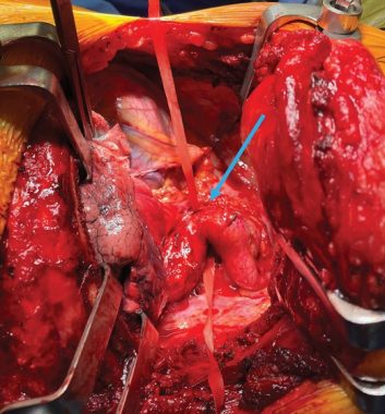
Image
Coarctation of the aorta (December 2022 [2])
Aortic coarctation is a localized narrowing in the thoracic aorta which is rarely diagnosed in adulthood. This image shows an aortic coarctation in a 70-year-old male. This was approached by left thoracotomy. The segment of coarctation was successfully resected while on the left-sided cardiac bypass and replaced by an interposition graft.
View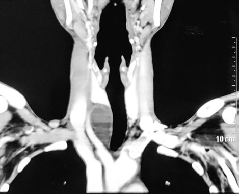
Image
Parathyroid cyst: enigma (December 2022 [1])
A 25-year-old gentleman, with chronic calcific pancreatitis and bilateral medullary nephrocalcinosis caused by hypercalcaemia, was found to have a 3.2×3.6 cm right inferior parathyroid adenoma. Contrast-enhanced computed tomography of the neck showed an enlarged well-defined cystic right inferior parathyroid gland located posterior to the right lobe of the thyroid with retrosternal extension. He underwent focused parathyroidectomy under the cervical block with the removal of a brown coloured cyst.
View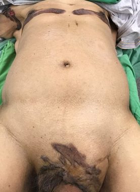
Image
Leser–Trélat sign (November 2022 [2])
An 80-year-old man presented as an emergency with a pneumoperitoneum. He was noted to have seborrhoeic keratoses on his chest bilaterally, his right axilla, and pubic regions that had developed in the month before admission. He was found to have metastatic sigmoid adenocarcinoma with liver metastases. The Leser–Trélat sign describes the eruption of multiple seborrhoeic keratoses secondary to disseminated malignancy.
View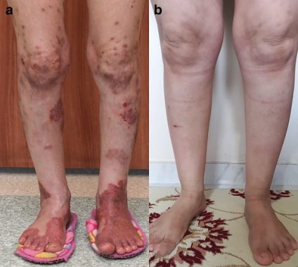
Image
Glucagonoma syndrome (November 2022 [1])
A 37-year-old female patient was referred with a widespread itchy rash, weight loss, and new-onset diabetes mellitus. Blood glucagon hormone measurement was >500 pg/ml. Laparoscopic distal pancreatectomy and splenectomy was performed. Her complaints disappeared afterwards. Surgery is the mainstay of treatment for glucagonoma disease and associated symptoms.
View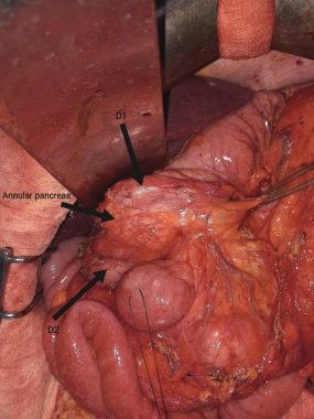
Image
Annular pancreas (October 2022 [2])
A 38-year-old man with a history of recurrent pancreatitis of undetermined origin was re-admitted with vomiting and epigastric pain. A decision was made to perform a laparoscopic cholecystectomy. At the laparoscopy, pancreatic tissue completely wrapped around the duodenum was found. A gastrojejunostomy was performed to treat the gastric outlet obstruction caused by the obstructive annular pancreas.
View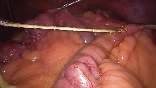
Image
Taenia saginata: unexpected finding during laparoscopic Roux-en-Y gastric bypass (October 2022 [1])
While performing the jejuno-jejunal anastomosis during a laparoscopic Roux-en-Y gastric bypass to treat morbid obesity, a yellowish filamentous structure appeared from the bowel lumen. It was removed, and subsequent microbiological analysis reported it to be a Taenia saginata (a tapeworm).
View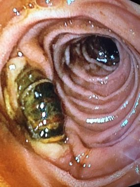
Image
Duodenal fistula of an aorto-mesenteric vascular bypass graft (September 2022 [2])
A 66-year-old man who had recently undergone aorto-mesenteric bypass presented with haematemesis. An upper gastrointestinal endoscopy found that the graft had fistulated into the lumen of the duodenum.
View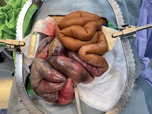
Image
Bowel ischaemia in encapsulating peritoneal sclerosis (September 2022 [1])
A 50-year-old male receiving peritoneal dialysis was admitted with abdominal pain. At exploratory laparotomy, small bowel ischaemia was found. In addition, the remaining small bowel was brown, hard, and like a plastic hose in keeping with early-stage encapsulating peritoneal sclerosis.
View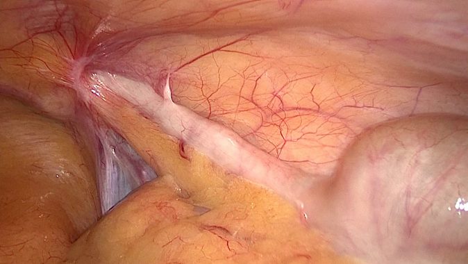
Image
Amyand’s hernia (August 2022 [2])
During a laparoscopic right inguinal hernia repair, the patient’s appendix was found in an indirect hernia sac. This finding is called an Amyand’s hernia.
View
The Swiss Society of Surgery was founded in 1913 as Swiss Society (hereafter SGC) for surgery.
Image
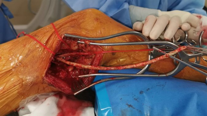
Long intact plaque extracted by femoral artery endarterectomy (August 2022 [1])
This intraoperative image shows an intact calcified plaque removed by endarterectomy. The plaque had caused a long occlusion of the superficial femoral artery. Superficial femoral artery endarterectomy should be considered when the saphenous vein is unavailable.
View

