
Snapshot quiz 13/31
Published: 07/26/2013
Authors: Barber Z, Di Souza N and Lintott P
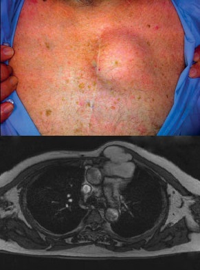
An 82-year-old man had an 18-month history of a slowly enlarging, painless subcutaneous lump on his chest wall (photograph). His past medical history included coronary artery bypass surgery. Magnetic resonance imaging identified a 7·2 × 4·3-cm aneurysmal dilatation of a vein bypass graft, extending through the intercostal space. Treatment options of surgical or radiological intervention were excluded when coronary angiography showed the vein graft to be patent and perfusing a significant proportion of the remaining functional myocardium. The patient was managed conservatively and follow-up imaging showed that the aneurysm had thrombosed spontaneously.