
Snapshot quiz 16/14
Published: 01/16/2017
Authors: Vashishtha N, Singhal D and Tandon R
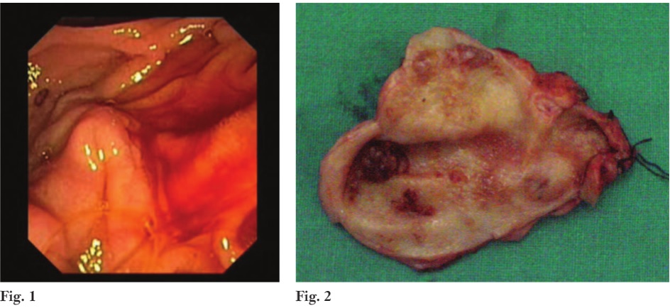
A 50-year-old woman presented with anaemia. Side-viewing endoscopy showed bleeding from the major duodenal papilla, diagnostic of haemobilia (Fig. 1). MRI showed a focal-enhancing lesion in the gallbladder fundus. The patient underwent cholecystectomy with frozen-section analysis followed by lymph node clearance. The surgical specimen (Fig. 2) revealed a friable polypoidal tumour at the fundus, which was a papillary adenocarcinoma (pT1a N0 Mx) on histopathology. Papillary gallbladder carcinoma may present rarely with anaemia due to haemobilia. The haemobilia is usually intermittent owing to periodic sloughing and therefore more difficult to diagnose.