
Snapshot quiz 16/4
Published: 06/06/2016
Authors: Agnes A, Biondi A, Ricci R, Riccioni M and Persiani R
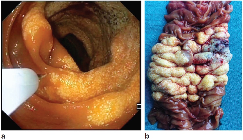
A 55-year-old woman presented with a 12-month history of iron deficiency anaemia and abdominal pain. Oesophagogastroduodenoscopy and colonoscopy were negative, whereas videocapsule enteroscopy identified an endo-luminal jejunal mass. CT showed thickening of the jejunal wall without contrast enhancement. Push enteroscopy confirmed the presence of a proximal jejunal stenosis, caused by a lesion presenting a yellowish-white and foamy appearance of the mucosa, with enlarged submucosal veins (a). An endoscopic tattoo was placed adjacent to the lesion, and the patient underwent a laparoscopic segmental resection of the jejunum. The resected specimen consisted of a 10-cm jejunal segment, hosting a tumour involving the entire visceral circumference (b). The final diagnosis was cavernous lymphangioma of the small bowel: a rare, benign, hamartomatous lesion that may occur in the mesentery, retroperitoneum and visceral organs.