
Splenic infarction
Published: 03/02/2012
Authors: Taylor TR and Lobo DN
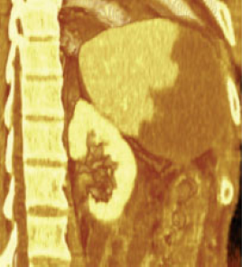
A 45-year-old man developed left-sided abdominal pain and pyrexia 3 weeks after a spleen-preserving distal pancreatectomy for chronic pancreatitis. Images obtained in the portal venous phase of a contrast-enhanced abdominal CT demonstrated thrombosis of the splenic vein; subsequent volume rendering of the CT images in an oblique coronal plane demonstrated a normally perfused superior portion of the spleen, with infarction of the inferior portion. The patient had a splenectomy and the line of demarcation on the resected specimen matched the imaging.