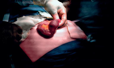
Leser–Trélat sign (November 2022 [2])
An 80-year-old man presented as an emergency with a pneumoperitoneum. He was noted to have seborrhoeic keratoses on his chest bilaterally, his right axilla, and pubic regions that had developed in the month before admission. He was found to have metastatic sigmoid adenocarcinoma with liver metastases. The Leser–Trélat sign describes the eruption of multiple seborrhoeic keratoses secondary to disseminated malignancy.
View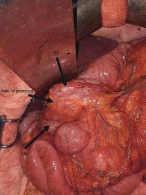
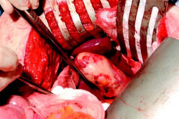
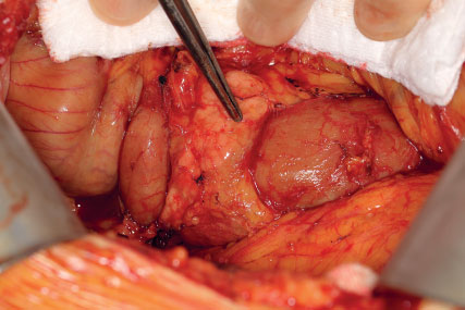
Annular pancreas (October 2022 [2])
A 38-year-old man with a history of recurrent pancreatitis of undetermined origin was re-admitted with vomiting and epigastric pain. A decision was made to perform a laparoscopic cholecystectomy. At the laparoscopy, pancreatic tissue completely wrapped around the duodenum was found. A gastrojejunostomy was performed to treat the gastric outlet obstruction caused by the obstructive annular pancreas.
View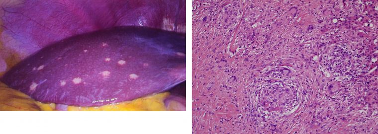
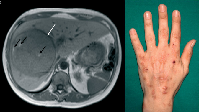
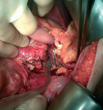
Unfamiliar liver lesions (May 2022 [1])
A 22 year-old male patient underwent laparoscopic cholecystectomy for recurrent biliary colic. Multiple macroscopic pale lesions were noted on the liver surface. Histopathology confirmed non-caseating granulomas. Serum angiotensin-converting enzyme was elevated. These findings were suspicious for extrapulmonary manifestations of sarcoidosis. Up to 10 per cent of all liver biopsy specimens are granulomas. Of these, 22 per cent are due to sarcoidosis. This is mostly asymptomatic. Up to 40 per cent of patients however present with biochemical derangement or hepatomegaly.
View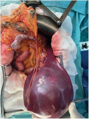
Snapshot quiz (January 2022 [2])
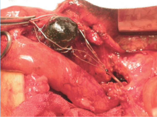
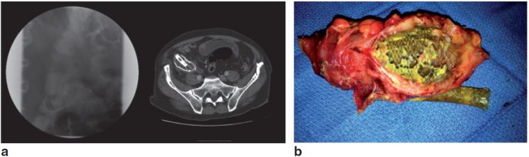
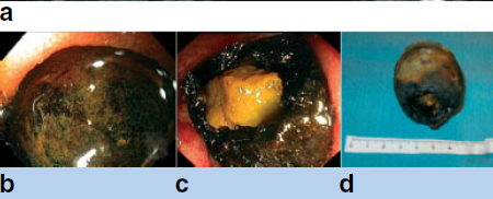
A 17-year-old woman presented with a 3-year history of an abdominal mass that was increasing in size. On abdominal examination there was a 20-cm mobile pelvic mass. What is shown in the intraoperative photograph?
View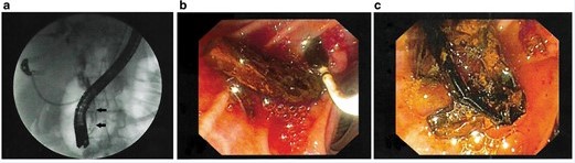
Snapshot quiz (October 2021 [1])
A 60-year-old woman underwent emergency therapeutic endoscopic retrograde cholangiopancreatography for three episodes of right hypochondriac pain, fever, and jaundice. The following findings were demonstrated on endoscopic views and on the cholangiogram. What is the diagnosis?
View
Snapshot quiz (June 2021 [2])
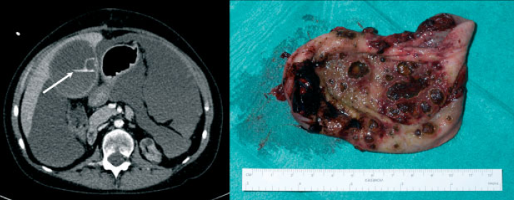
A 69-year-old male who initially presented with Streptococcus bacteremia was later readmitted with recurrent fevers, chills, and abdominal pain. A contrasted computed tomography (CT) scan of the abdomen and pelvis was obtained. What is the diagnosis and management?
View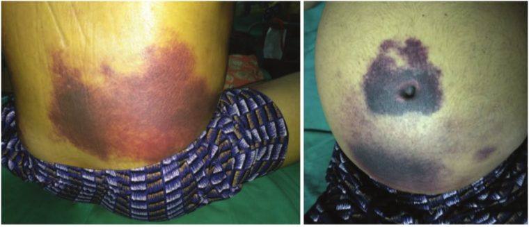
Snapshot quiz 18/9
A 45-year-old alcoholic man presented with acute epigastric pain spreading across the whole abdomen. What is the likely cause?
View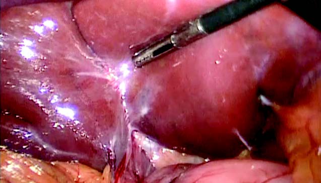
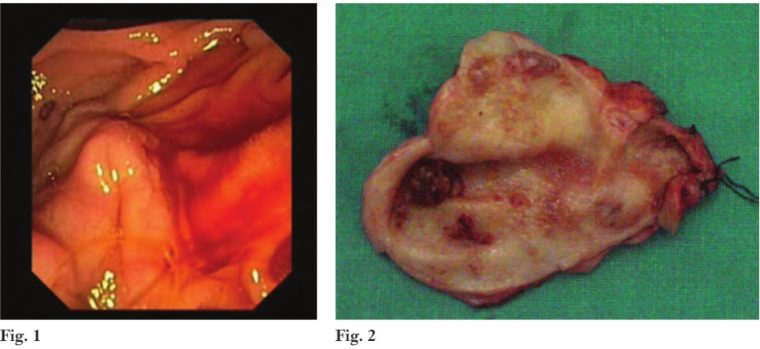
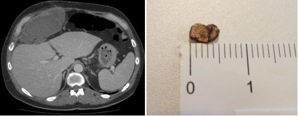
Snapshot quiz 16/14
What is happening to the ampulla of Vater on endoscopy (Fig.1), and what is the cause, which was later removed at operation (Fig.2)?
View
Snapshot quiz 14/9
This 65-year-old man was treated for gastric outlet syndrome and jaundice due to pancreatic cancer with two metallic stents. What has happened?
View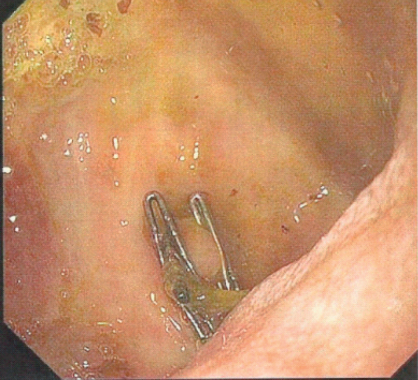
Snapshot quiz 13/29
Snapshot quiz 13/28
Snapshot quiz 13/23
Snapshot quiz 12/18
Snapshot quiz 12/13
Snapshot quiz 11/11
Snapshot quiz 11/6
Snapshot quiz 11/5
The diagnostic value of silver stool (Thomas’ sign)
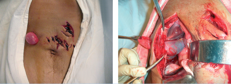
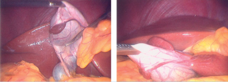
Partial gallbladder prolapse following abdominal stab injury
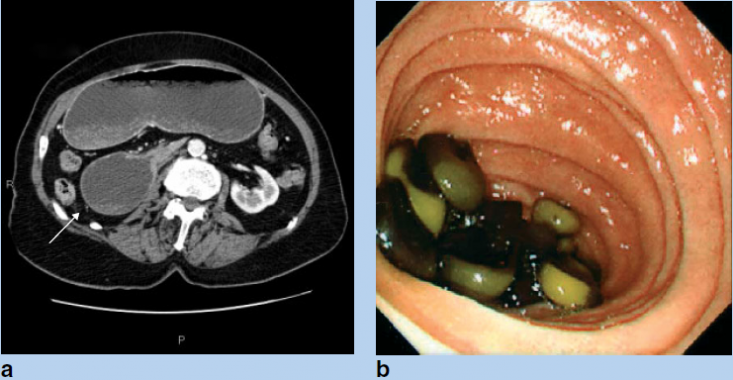
Multiple duodenal gallstones found at endoscopy
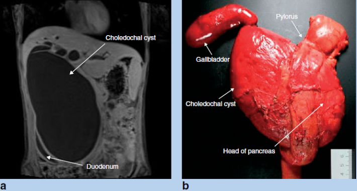
Giant choledochal cyst
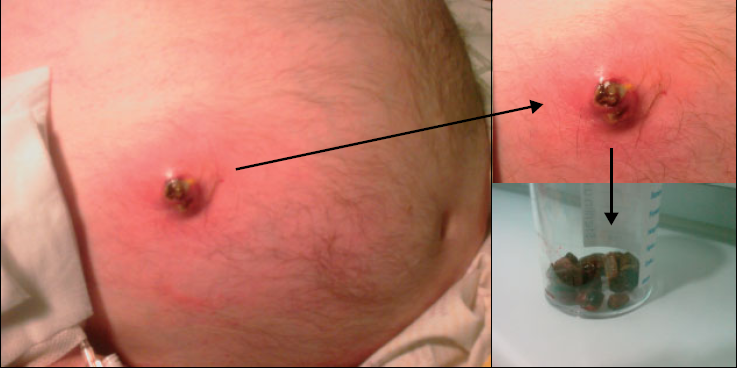
Gallstone impacted in the skin of the abdominal wall
Gall bladder associated ectopic liver
Cytomegalovirus cholecystitis
Bouveret’s Syndrome