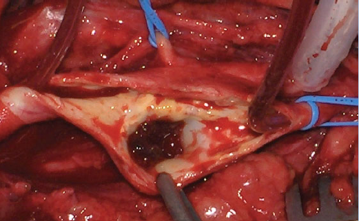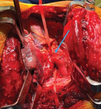
Coarctation of the aorta (December 2022 [2])
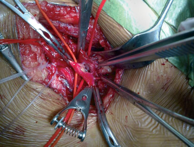
Aortic coarctation is a localized narrowing in the thoracic aorta which is rarely diagnosed in adulthood. This image shows an aortic coarctation in a 70-year-old male. This was approached by left thoracotomy. The segment of coarctation was successfully resected while on the left-sided cardiac bypass and replaced by an interposition graft.
View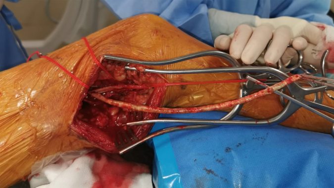
Long intact plaque extracted by femoral artery endarterectomy (August 2022 [1])
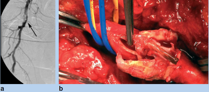
This intraoperative image shows an intact calcified plaque removed by endarterectomy. The plaque had caused a long occlusion of the superficial femoral artery. Superficial femoral artery endarterectomy should be considered when the saphenous vein is unavailable.
View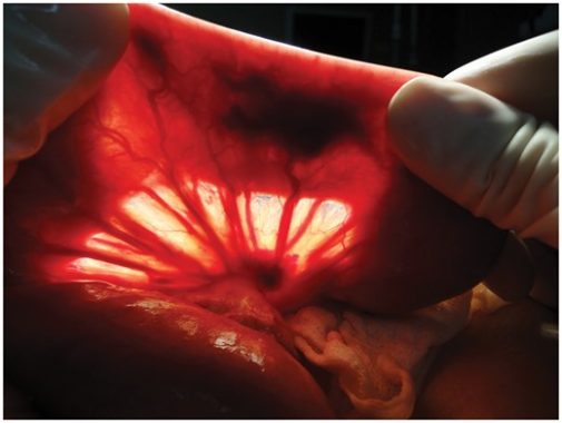
Snapshot quiz (November 2021 [2])
A 72-year-old man presented with recurrent attacks of melaena and severe anaemia. Oesophagogastroduodenoscopy and colonoscopy were normal. Contrast-enhanced abdominal CT showed extravasation of contrast medium in the left abdomen. Ongoing bleeding necessitated surgical intervention. A 1.5 × 2.0-cm lesion in the proximal jejunum, 10 cm distal to the Treitz ligament, was found at surgery. What is the diagnosis?
View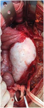
Snapshot quiz (November 2021 [1])
A 65-year-old man presented to the emergency department with chronic abdominal pain. His medical history included hypertension, tobacco use, and he had lost 8 kg during the previous 6 months. His physical examination was notable for a distended abdomen with a pulsatile mass at the epigastrium. Laboratory tests showed elevated inflammatory markers. He underwent surgery. What is shown in the intraoperative picture?
View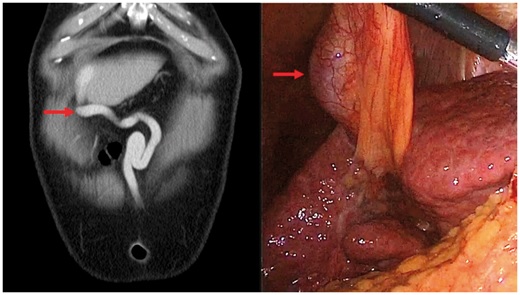
Snapshot quiz (March 2021 [2])
A 70-year-old man with chronic liver disease was diagnosed with colonic adenocarcinoma. What is shown, and what is the cause of the abnormality seen on the coronal image taken from the staging CT scan and on the intraoperative laparoscopic image?
View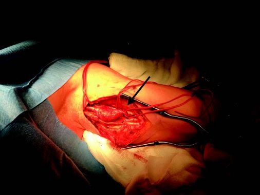
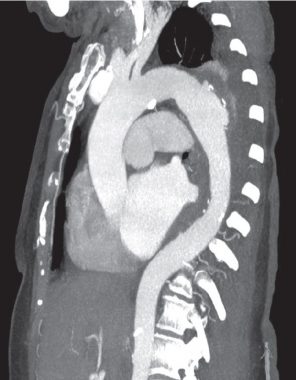
Snapshot quiz 18/3
A 28-year-old man was hit by a car while travelling on his scooter at 30 m.p.h. On initial assessment in the accident and emergency department he had multiple fractures. The CT scan below was taken as part of a trauma series. What is the diagnosis, and how should the condition be treated?
View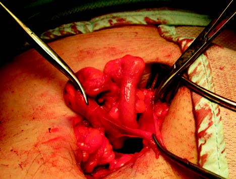
Snapshot quiz 17/3
This was the finding at elective right groin lymph node biopsy. What is the diagnosis?
View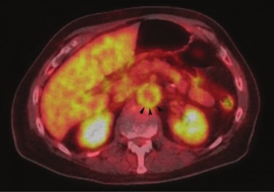
Snapshot quiz 16/2
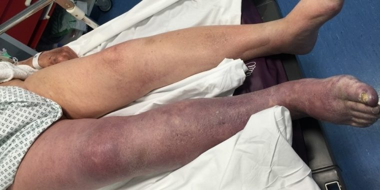
This man has a painful swollen leg with a palpable foot pulse. What is the diagnosis?
View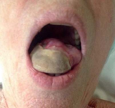
Snapshot quiz 15/5
A 74-year-old woman presented to the emergency department with a painful ulcerated necrotic lesion on the right side of her tongue. She had a 2-week history of new-onset headache, blurred vision and jaw claudication. What is the diagnosis and how could it be confirmed?
View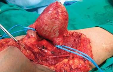
Snapshot quiz 14/4
This 37-year-old woman with end-stage renal disease underwent right-sided brachiocephalic arteriovenous fistula formation 8 years previously. She noticed a swelling in the right elbow region 2 years after the operation, which rapidly increased in size (intraoperative photograph shown). What is this condition and how should it be treated?
View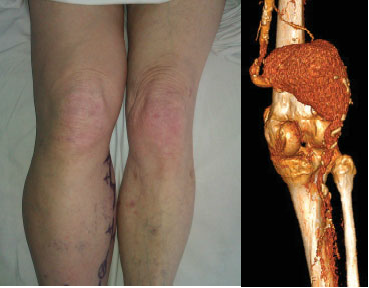
Snapshot quiz 13/36
Snapshot quiz 13/33
Snapshot quiz 13/31
Snapshot quiz 12/16

Snapshot quiz 12/14

Snapshot quiz 12/11

Snapshot quiz 12/8b
Snapshot quiz 12/4

Snapshot quiz 12/1
Snapshot quiz 11/13
Snapshot quiz 11/9
Arteriovenous fistula following endovenous laser treatment
Unusual cause of claudication

Prominent superficial temporal artery and transient ischaemea
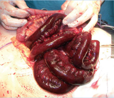
Mesenteric thrombosis
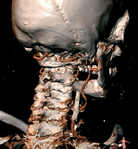
Carotid stent occlusion with persistent flow
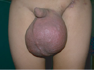
Pulsatile scrotum
