
Image
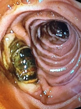
Duodenal fistula of an aorto-mesenteric vascular bypass graft (September 2022 [2])
A 66-year-old man who had recently undergone aorto-mesenteric bypass presented with haematemesis. An upper gastrointestinal endoscopy found that the graft had fistulated into the lumen of the duodenum.
View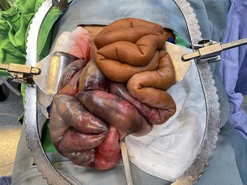
Image
Bowel ischaemia in encapsulating peritoneal sclerosis (September 2022 [1])
A 50-year-old male receiving peritoneal dialysis was admitted with abdominal pain. At exploratory laparotomy, small bowel ischaemia was found. In addition, the remaining small bowel was brown, hard, and like a plastic hose in keeping with early-stage encapsulating peritoneal sclerosis.
View
Image
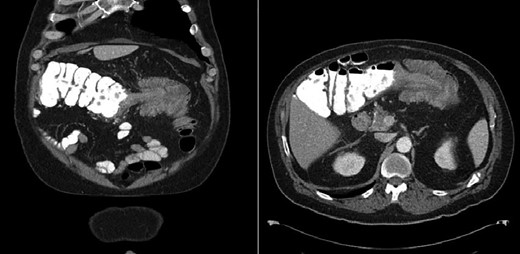
Adult colo-colonic intussusception (June 2022 [2])
A 62-year-old patient presented with multiple episodes of haematochezia, abdominal pain, nausea, and vomiting. Computed tomography demonstrated an 11 cm colo-colic intussusception with a lead point. Intraoperative and pathological findings found a benign 8.2 cm polypoid submucosal lipoma with ischaemic changes, ulceration, and necrosis of the surrounding colonic mucosa.
View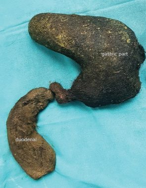
Image
Gastroduodenal trichobezoar: rare case of Rapunzel syndrome (May 2022 [2])
A 23-year-old woman presented with symptoms of obstruction. At laparotomy a gastroduodenal trichobezoar was removed from her stomach and duodenum. Trichobezoar are concretions of swallowed hair bundles and can be associated with Rapunzel syndrome. Endoscopy may be helpful in early cases; however, surgery remains the mainstay for late presentation.
View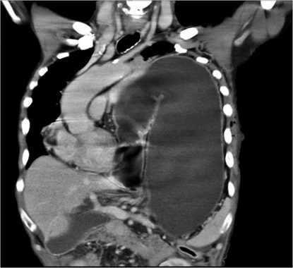
Image
Snapshot quiz (April 2022 [2])
A 76-year-old male with a known left diaphragmatic hernia presented with acute shortness of breath, vomiting and diarrhoea for five days. A chest computed tomography was performed. What is the diagnosis?
View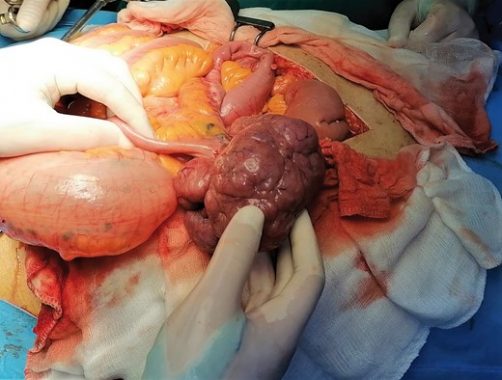
Image
Snapshot quiz (April 2022 [1])
A 55-year-old man presented acutely with small bowel obstruction. What is shown in the intraoperative photograph?
View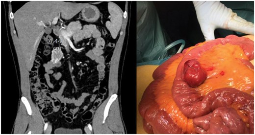
Image
Snapshot quiz (December 2021 [1])
A 37-year-old, previously healthy, man presented with profuse rectal bleeding necessitating transfusion. Colonoscopy and oesophagogastroduodenoscopy were normal. CT angiography was performed. What is the diagnosis?
ViewScientific Surgery
Authors: Wu L, Shang R, Sharma P, Zhou W, Liu J, Yao L et al.
Scientific Surgery
Authors: Ojima T, Nakamura M, Hayata K, Kitadani J, Katsuda M, Takeuchi A et al.
Scientific Surgery
Authors: Yang H, Liu H, Chen Y, Zhu C, Fang W, Yu Z et al.
Scientific Surgery
Authors: Van Workum F, Verstegen MHP, Klarenbeek BR, Bouwense SAW, van Berge Henegouwen MI, Daams F et al.
Scientific Surgery
Authors: Lee J, Garvey EM, Bundrant N, Hargis-Villanueva A, Kang P, Osuchukwu O et al.
Scientific Surgery
Authors: Wang H, Tang H, Fang Y, Tan L, Yin J, Shen Y et al.
Scientific Surgery
Authors: O’Leary DP, Walsh SM, Bolger J, Baban C, Humphreys H, O’Grady S et al.
Scientific Surgery
Authors: Nuytens F, Dabakuyo-Yonli TS, Meunier B, Gagnière J, Collet D, D’Journo XB et al.
Scientific Surgery
Authors: Adamson DA, Byrne A, Porter C, Blazeby J, Griffiths G, Nelson A et al.
Image
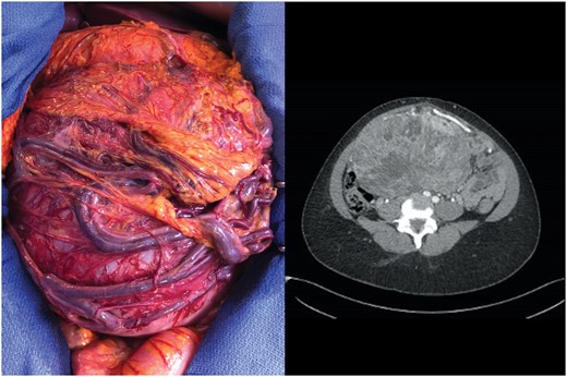
Snapshot quiz (August 2021 [2])
A 34-year-old woman presented with a 20 cm intra-abdominal tumour with large draining veins and shunting surrounding the mass, detected by abdominal computed tomography (CT) scan. Six years before she underwent surgery for a pelvic tumour. What is the diagnosis?
View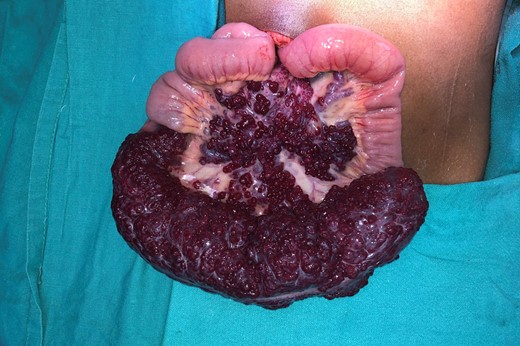
Image
Snapshot quiz (August 2021 [1])
An 8 year old boy presented with colicky abdominal pain and chronic anemia. Faecal occult blood analysis was positive, upper and lower GI endoscopy were unremarkable. 99mTC labeled RBC scan showed an abnormal accumulation of tracer in the distal small bowel on the right side of abdomen. Diagnostic laparoscopy followed by open resection was done. What is the diagnosis?
View